Document Type : Original Research Article
INTRODUCTION
The application of nanotechnology products in human activities is increasing extensively. Nanoparticles are widely used in industry, electronics, and medicine [1]. Cobalt is a magnetic mineral, and the cobalt nanoparticle has special properties, including a large surface area that is associated with its small size and its catalytic and magnetic properties [2]. Cobalt nanoparticles are widely used in industrial applications such as chemical catalysts, gas sensitive equipment, coatings, optical reagents, magnetic strips, as well as in medical biotechnology, including magnetic resonance images and experimental cancer treatments [4,5].
Ag NPs are used in a variety of fields, including food, medicine, and industrial purposes, which is associated with its chemical and physical properties [6]. These properties of Ag NPs include biological properties, high electrical conductivity, thermal and optical properties [7-8]. Due to its properties, Ag NPs have several uses, including as antimicrobial agents, in industry, household appliances, health care products, consumer products, medical devices coverage, optical sensors, in the pharmaceutical industry, food industry, orthopedics, and as anticancer agents [9].
In a study by Bocate and colleagues in 2019, it has been reported that Ag NPs and simvastatin have anti-fungal activity against toxigenic species of Aspergillus [10]. The Ag NPs have antibacterial activity against Pseudomonas aeruginosa [11]. In a study by Li and colleagues in 2007, it has been reported that the Ag NPs exhibit antimicrobial activity against Staphylococcus aureus and Escherichia coli [12]. Ag NPs have antifungal activity against Candida spp [13]. Ag NPs have inhibitory effects against bovine herpesvirus-1[14].
Ag NPs have anti-inflammatory activity in rats [15]. In addition, it has been shown that Ag NPs possess antimicrobial properties, reduce inflammation and modulate the fibrogenic cytokines [16]. A recent study have shown that Ag NPs have anti-angiogenic properties [17]. It has been proven that Ag NPs not only induce planned cell death, but are also sensitive to cancerous cells [17]. Ag NPs induce changes in cellular morphology and increase oxidative stress leading to mitochondrial damage [18].
Exposure to cobalt nanoparticles causes oxidative stress, inflammation and DNA injury [19,20]. Generally, some mineral nanoparticles may have effects on carcinogens and cellular poisoning that are related to the chemical nature of minerals. Nanoparticles can cause DNA damage and oxidative stress through the production of ROS [21]. Exposure to cobalt nanoparticles induces lung injury and DNA mutations in mice [22]. However, some studies have shown the effects of genetic poisoning on some of the nanoparticles, and their mechanism is still unclear. In vitro studies have shown that exposure to cobalt nanoparticles leads to DNA damage and cellular deformation, in which ROS and oxidative stress are involved [23,24].
Considering the side effects of AgCo NPs in this study, the effects of different amounts of AgCo NPs on the levels of AST, ALT, ALP, ALP, albumin and total protein and liver tissue changes in adult male rats were investigated. It is necessary to be cautious about functional tests and liver tissue changes.
MATERIALS AND METHODS
Synthesis of AgCo NPs
Silver-cobalt nanoparticles were synthesized by two step process. At first, silver nanoparticles were prepared by electrochemical method. For this purpose, a solution of electrolyte was containing Silver nitrate, Potassium nitrate and Polyvinylpyrrolidone. Silver nanoparticles were synthesized during 75 and 300 seconds. During the experiment, the cathode electrode was rotating with 3000 rpm of speed and current was 1 A. At second step, a solution of Cobalt sulfate and Cetyl trimethylammonium bromide was added to the same amount of silver nanoparticles which synthesized from the first step. Then, for preparation of silver-cobalt nanoparticle; Sodium borohydride was added drop by drop as a reducing agent. Consequently, the color change of the solution to dark brown indicated the formation of silver-cobalt nanoparticles by reduction method. All these experiments were done at room temperature [25].
Animals
In this experimental study, 28 male Wistar rats weighing 180-220 g and age 2.5 to 3 months were used. Animals were randomly divided into 4 groups of 7 animals until the test was performed in standard cages under the same conditions at 20- 22 ° C and with 12-hour light and 12-hour darkness. They were provided with enough water and food, and ethical considerations about animals were respected.
Animal treatment
The animals were divided into 4 groups of 7, which included: control group: were not affected by any drug treatment. Experimental group1 received 25mg / kg of AgCo NPs in which Ag NPs were synthesized during 75 seconds intraperitoneally for 14 days. Experimental group 2 received 100 mg / kg of AgCo NPs in which Ag NPs were synthesized during 75 seconds intraperitoneally for 14 days. Experimental group 3 received 25mg / kg of AgCo NPs in which Ag NPs were synthesized during 300 seconds intraperitoneally for 14 days. Intake doses and injection type and duration of injection were selected using previous studies [26-28].
After the end of the treatment period, animals were affected by anesthesia with ether. Blood collection from the left ventricle of the heart. The blood samples were kept in laboratory for 20 minutes and centrifuged for 15 minutes at 5000 rpm. Serum was separated from the clot. Measurements of AST, ALT and DGKC, ALP were performed using P-nitrophenyl phosphate AMP method. The biuret reaction end point method was used to measure total protein. In this experiment, protein in the alkaline environment with copper ions and tartrate causes the formation of azure color, and the color intensity created is proportional to the total amount of protein in the sample. Bromocresol Green was used to measure albumin in which bromocresol and albumin produced a colored complex. The intensity of the color created is proportional to the amount of albumin in the sample [29,30].
Histological experiments
After the autopsy, the animal liver was removed. At the stage of fixation, tissues were established in 10% formaldehyde-formaldehyde. The dehydration step was carried out with the aid of alcohol with different concentrations (low to high). The clarification step was carried out by placing tissues in two containers containing Xylan. In the stage of tissue replacement in three dishes containing molten paraffin (65 ° C), each one was placed in an hour. In the molding stage, the paraffin embedded sample is placed inside with a mold filled of molten paraffin. In the cross section, the sections were cut to a thickness of 4.5 microns. The staining stages of hematoxylin-eosin paint were used. All histological studies were conducted under the supervision of the pathologist [31].
Statistic analysis
Data were analyzed using SPSS-22, ANOVA and Duncan test. The statistical inference line was used to examine the significant difference between the experimental groups receiving different amounts of AgCo NPs at the level of P <0.05 compared with the control group. In this research, the results of the experiments carried out along with the corresponding statistical calculations are presented in the table.
RESULTS AND DISCUSSION
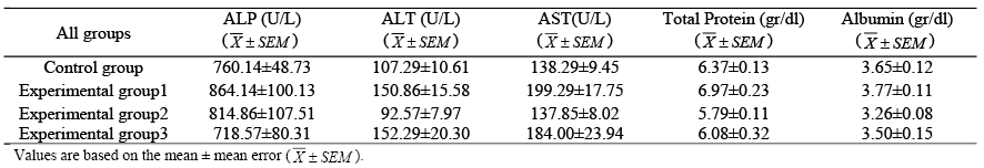
In this study, statistical analysis and comparison of mean serum levels of AST, ALT, ALP, albumin and total protein in the experimental groups receiving AgCo NPs were in comparison with the control group. P <0.05 was the basis for statistical inference to examine the significant difference between different groups.
The mean serum level of aspartate aminotransferase (AST) enzyme in the experimental groups receiving AgCo NPs did not significantly change in compared to the control group (P <0.05) (Table 1).
The mean serum level of alanine aminotransferase (ALT) in the experimental groups receiving AgCo NPs did not significantly change significantly in compared to the control group (P <0.05) (Table 1).
The mean serum level of alkaline phosphatase (ALP) in the experimental groups receiving AgCo NPs did not change significantly in compared to the control group (P <0.05) (Table 1).
The mean serum albumin level in the experimental groups receiving AgCo NPs did not significantly change in comparison with the control group (P <0.05) (Table 1).
The mean serum total protein level in the experimental groups receiving AgCo NPs did not significantly change in comparison with the control group (P <0.05) (Table 1).
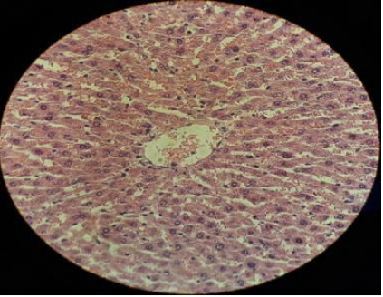
The results of liver histological studies showed that liver tissue in the control group was completely normal and without cell damage. This group has a cellular structure and maintains the radial and natural state of the liver cells (a nucleus and two nuclei) and the presence of the nucleus and central vein of its distinctive features (Fig. 1).
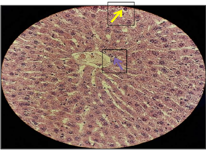
The liver tissue in the experimental group 1 is completely normal and does not have cell damage. This group has a cellular structure and maintains the radial and natural state of the liver cells (a nucleus and two nuclei) and the presence of the nucleus and central vein of its distinctive features (Fig. 2).
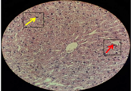
The liver tissue in the experimental group 2 is completely normal and does not have cell damage. This group has a cellular structure and maintains the radial and natural state of the liver cells (a nucleus and two nuclei) and the presence of the nucleus and central vein of its distinctive features (Fig. 3).
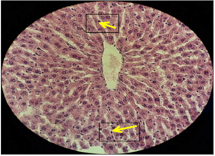
The liver tissue in the experimental group 3 is completely normal and does not have cell damage. This group has a cellular structure and maintains the radial and natural state of the liver cells (a nucleus and two nuclei) and the presence of the nucleus and central vein of its distinctive features (Fig. 4).
According to the results of the present study, serum levels of aspartate aminotransferase (AST) enzymes, albumin and total protein did not change significantly in the experimental groups receiving AgCo NPs compared to the control group. Mean serum alanine aminotransferase (ALT) and alkaline phosphatase levels (ALP) in experimental groups receiving AgCo NPs did not change significantly compared to the control group. In experimental groups receiving silver-cobalt nanoparticles, the liver tissue was healthy and unchanged.
The core of the AgCo NPs is composed of Ag NPs. Recent findings in in vitro studies suggest that Ag NPs can penetrate into cell membranes and other biological dams [32]. Ag NPs can inactivate intracellular organs, including mitochondria, which impair the membrane potential and thus induce the production of reactive oxygen species (ROS). With ROS accumulation, oxidative stress increases, followed by cellular poisoning with Ag NPs [33].Liver cells, according to the need for ATP, are the bulk of their volunteers for liver toxicity induced by Ag NPs, reported in in vivo studies. A study in Invivo proved that Ag NPs induced liver toxicity by reducing ATP and reducing the activity of antioxidant enzymes, especially glutathione. Thus, cells treated with Ag NPs have abnormal shape and size [34].
Also, in the study of Recordati et al. In 2015, the induction of the smallest Ag NPs (10 nm) stimulated the distribution of silver tissue and liver-bile duct toxicity compared to larger silver nanoparticles (100 and 40 nm). This study showed that the effects of silver nanoparticles are dependent on size [35].Additionally, in a study by Xue et al in 2016, the Ag NPs induced cell death and induced cellular poisoning in HepG2 liver cells in humans. Exposure to silver nanoparticles can cause disturbances in the G2 / M phase and significantly increase the ratio of planned cell death and ROS production and reduce MMP in HepG2 cells. These results showed that the Ag NPs cellular mechanism may be related to oxidative stress induced by ROS production, leading to mitochondrial damage and induced cell death [36].
In a study by Shivastava et al in 2016, exposing mice to Ag NPs induced oxidative stress. Exposure to Ag NPs increases the symptoms of inflammation and interleukin-6 and nitric oxide synthase, which indicates liver toxicity [37]. Also, in a study by Lee et al in 2013, it was shown that Ag NPs were induced by autophagy. In addition, Ag NPs cause bioenergic defects associated with autophagy and cellular death in the rat liver. In this study, exposure to Ag NPs in in vivo conditions was found to lead to liver toxicity [38].
In addition, in a study by Heydarnjad et al in 2015, showed that oral exposure to Ag NPs caused a lot of changes in blood biochemical factors and liver toxicity, including increased levels of ALT, AST, and liver tissue damage in mice treated with Ag NPs compared to control group [39]. Also, in a study by Zhu et al in 2016, The Ag NPs induces the planned cell death through ROS-mediated pathways in HepG2 cells [40].
In a study by Mendonça et al in 2018, the single dose of Ag NPs (5 mg/kg b.w.) caused liver toxicity through the accumulation of Ag NPs in the rat liver [41]. Also, in a study by Ahmadian et al in 2018, showed that Ag NPs could be used as a strong chemotherapy agent to treat hepatocellular carcinoma [42]. In a study by Moradi-Sardareh et al in 2018, Ag NPs (0.25 mg/kg) significantly altered oxidative stress in the serum and liver tissue, but did not change the serum levels of liver enzymes in mice [43]. Also, in a study by Cho et al in 2018, it was shown that intraperitoneal administration of 10nm Ag NPs led to vacuolation, single cell necrosis, and focal necrosis in the liver of mice [44].
The AgCo NPs coating is made up of cobalt nanoparticles. One study that focused on the genetic toxicity of cobalt ferrite nanoparticles in the rat liver proved that exposure to cobalt ferrite nanoparticles resulted in increased expression of axial genes associated with oxidative stress, planned cell death, DNA injury, and cellular damage [45].
Histologic findings have shown that exposure to cobalt nanoparticles can lead to more severe liver damage than cadmium chloride, however, serum symptoms show vague changes. Cobalt accumulates in the liver, kidneys, pancreas and heart. Salts and minerals cobalt induce oxidative damage to DNA through reactive oxygen species (ROS) , and inhibit DNA repair [46]. In the study of Garoui et al in 2011, liver toxicity was observed in exposed cobalt chloride rats [47].
The BCL2 family of proteins comprises the planned cell death anti-death proteins, including BCL-2 and pre-apoptotic Bax protein, which are central regulators of mitochondrial cell death. The BCL2 family proteins regulate the release of cytochrome C from mitochondria to cytosol. BCL-2 proteins stabilize cytochrome C release by protecting transmissive permeability and stabilizing mitochondrial membrane function, while Bax proteins induce cytochrome C release [48,49]. In addition, BCL-2 family proteins can regulate the production of ROS [48,49]. Increasing the ROS molecularly inhibits expression of BCL-2 and improves Bax expression. A study by Liu et al in 2016 showed that cobalt nanoparticles increase the ratio of Bax to BCL2 levels, and these results indicate that cobalt nanoparticles regulate the permeability of an outer mitochondrial membrane through BCL-2 family proteins, which results in the release of cytochrome C and ultimately leads to the planned cell death of BRL-3A cells [50].
The results of this study are not consistent with other researchers’ findings, and it seems that AgCo NPs did not cause liver toxicity due to low dosage and short duration of the test. Considering the results, it can be concluded that factors such as nanoparticle size, dosage, and nanoparticle coating play an important role in the effects of nanoparticles.
CONCLUSION
In general, the results of this research showed that AgCo NPs do not have adverse effects on functional and liver tissue tests in adult male rats. However, the study of the effects of other doses of AgCo NPs on the liver and other organs of the human body and animals is recommended.
ACKNOWLEDGEMENT
Hence, we sincerely appreciate the close cooperation of the Vice-Chancellor of Research at Shiraz Azad University.
CONFLICT OF INTEREST
The authors declare that there are no conflicts of interest regarding the publication of this manuscript.